🫀
Integumentary · Anatomy
Skin Layers
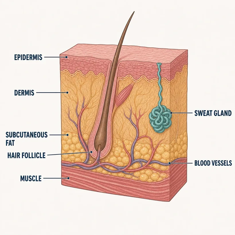
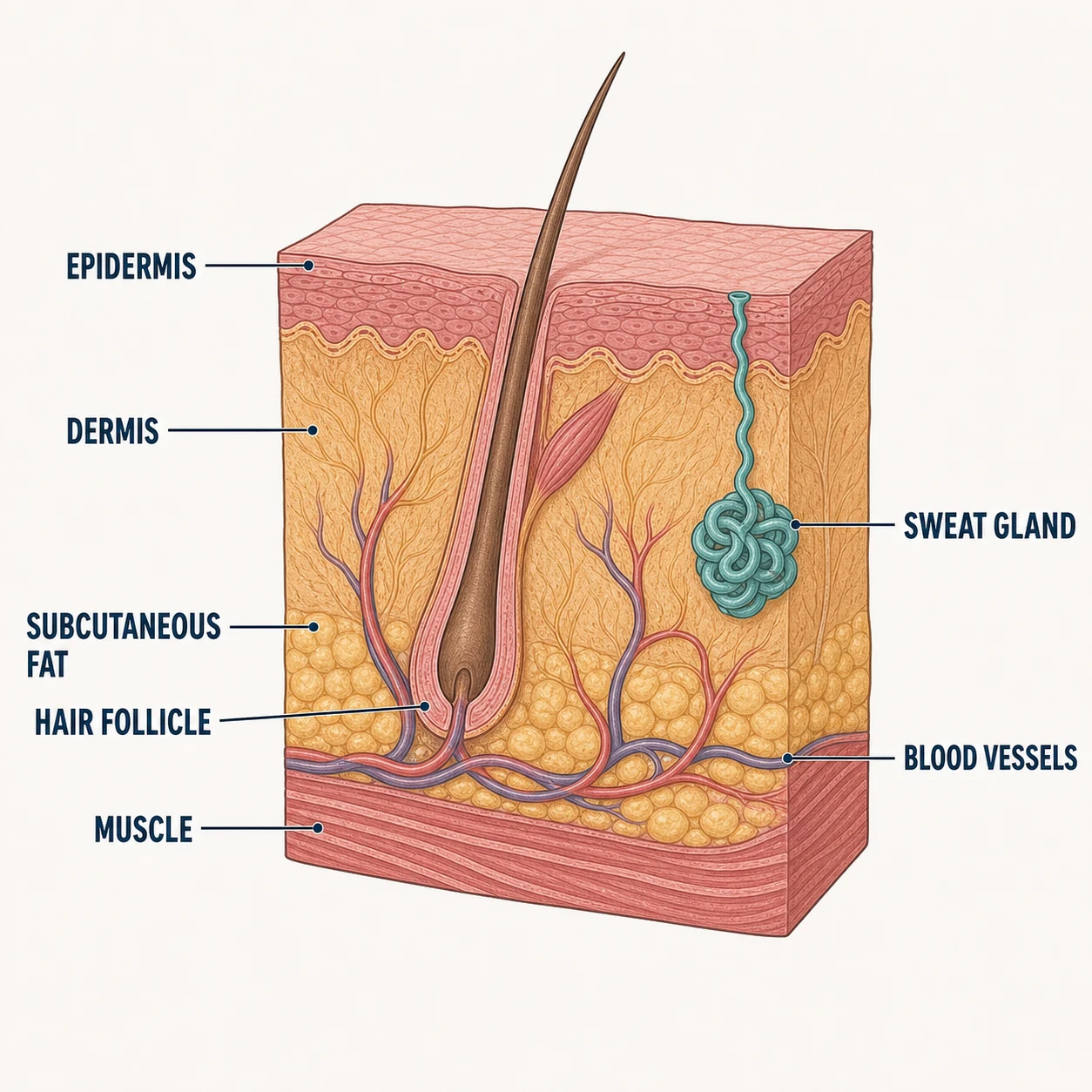
Epidermis, dermis, subcutaneous fat — pressure-injury staging is literally this diagram.
The structures
- Epidermis — Waterproof barrier; no blood vessels — Stage 1–2 territory
- Dermis — Vessels, nerves, follicles — partial-thickness wounds end here
- Subcutaneous fat — Insulation and cushioning — Stage 3 exposes it
- Fascia & muscle — Stage 4 reaches this deep
- Sweat glands — Thermoregulation
- Sebaceous glands — Oil; blocked ones become acne and cysts
Why it matters on shift
- Stage 1 is intact skin with non-blanchable redness — the only stage you can fully reverse; float that heel today
- Can't see the wound base under slough? It's UNSTAGEABLE, not Stage 4 — document what you see
- Deep tissue injury looks like a bruise on a pressure point — it will open, and documentation now protects you later
Source: Anatomical structures per OpenStax Anatomy & Physiology, 2nd ed. (openstax.org, CC BY 4.0). Educational reference — verify clinical application against your facility's policy.Reference only — verify against your facility's policy.