🫀
Renal · Anatomy
Kidney & Nephron
A million nephrons filter, reabsorb, and secrete — urine is what's left.
The structures
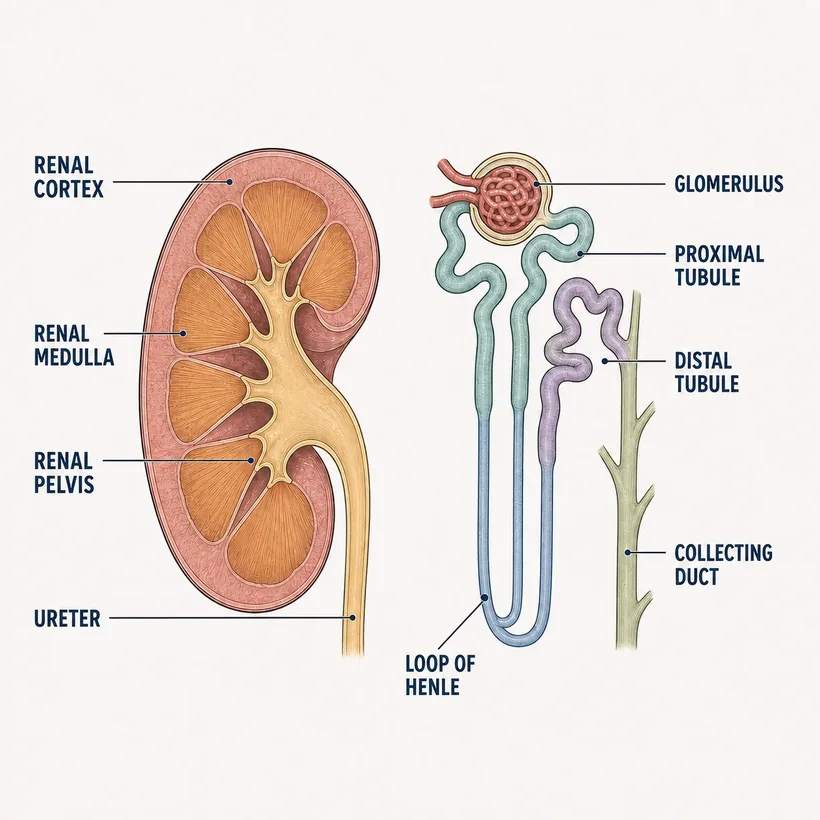
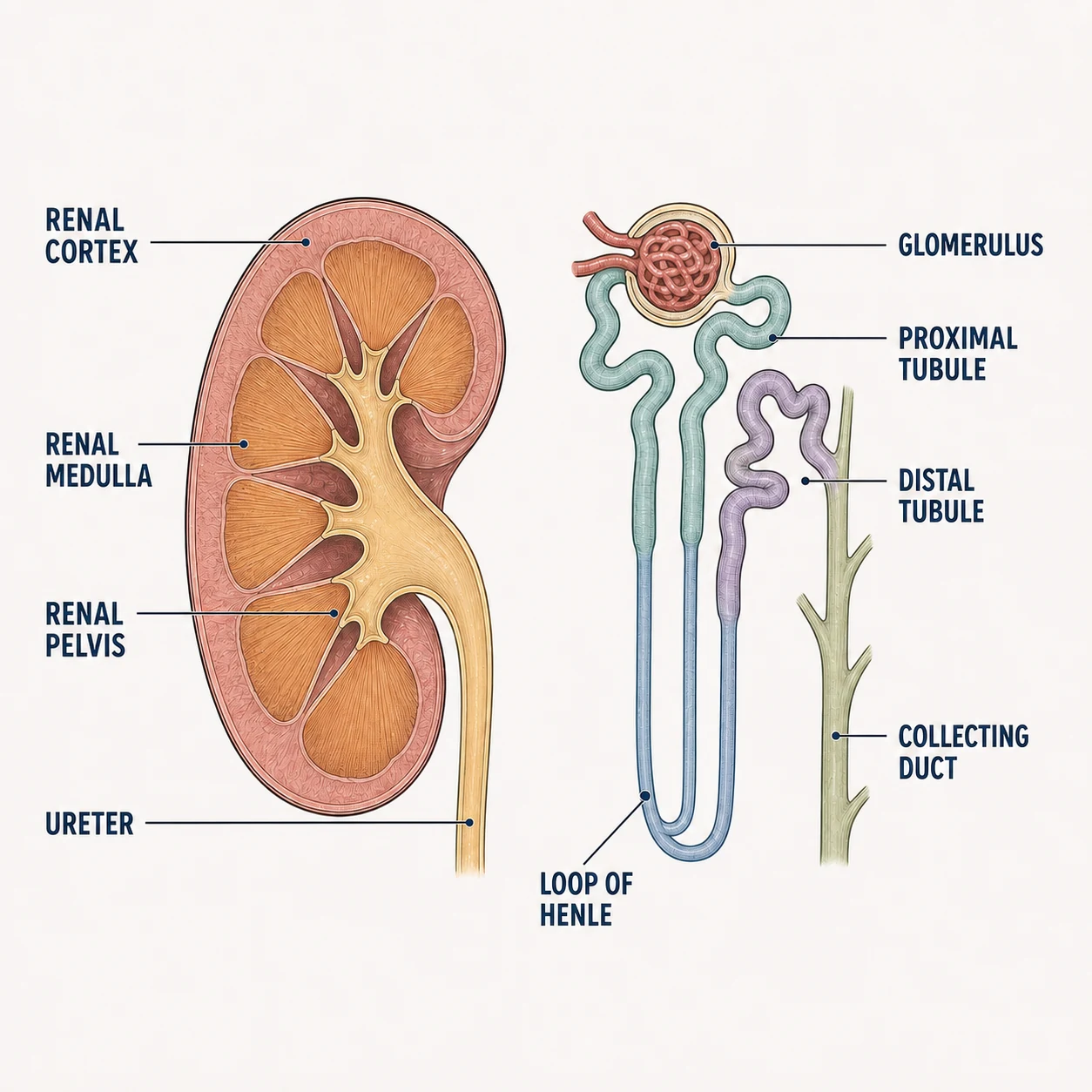
- Renal cortex — Outer layer — most glomeruli live here
- Renal medulla — Pyramids that concentrate urine
- Glomerulus — Filter head — where GFR happens
- Proximal tubule — Reabsorbs most of the good stuff (glucose, electrolytes)
- Loop of Henle — Concentration gradient — loop diuretics act here
- Distal tubule — Fine-tunes sodium and potassium — thiazides act here
- Collecting duct — ADH controls final water reabsorption
- Ureter — Urine to the bladder
Why it matters on shift
- Creatinine rising = glomeruli filtering less — recheck the med list for nephrotoxins (NSAIDs, contrast, vanco)
- Loop diuretics dump potassium; K-sparing ones hold it — know which your patient takes before you see the K result
- Oliguria < 0.5 mL/kg/hr is the number to report, not just 'low urine'
Source: Anatomical structures per OpenStax Anatomy & Physiology, 2nd ed. (openstax.org, CC BY 4.0). Educational reference — verify clinical application against your facility's policy.Reference only — verify against your facility's policy.