🫀
Sensory · Anatomy
Ear
Outer ear collects, middle ear amplifies, inner ear converts to nerve signal — and runs your balance.
The structures
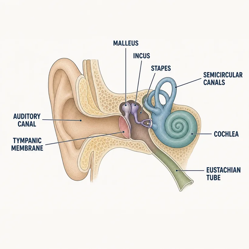
- External auditory canal — Sound in; cerumen made here
- Tympanic membrane — Eardrum — vibrates with sound
- Malleus, incus, stapes — Three tiny bones amplifying vibration
- Eustachian tube — Pressure equalizer to the pharynx — the otitis media highway in kids
- Cochlea — Hearing organ — hair cells convert vibration to signal
- Semicircular canals — Balance — vertigo lives here
- Vestibulocochlear nerve (CN VIII) — Carries hearing and balance to the brain
Why it matters on shift
- Gentamicin, vancomycin, furosemide are ototoxic — new tinnitus or hearing change on these meds is reportable
- True vertigo (room spinning) is an inner-ear or CN VIII problem — different workup than 'dizzy when I stand'
- Kids' eustachian tubes are short and horizontal — why ear infections follow every cold
Source: Anatomical structures per OpenStax Anatomy & Physiology, 2nd ed. (openstax.org, CC BY 4.0). Educational reference — verify clinical application against your facility's policy.Reference only — verify against your facility's policy.