🫀
Sensory · Anatomy
Eye
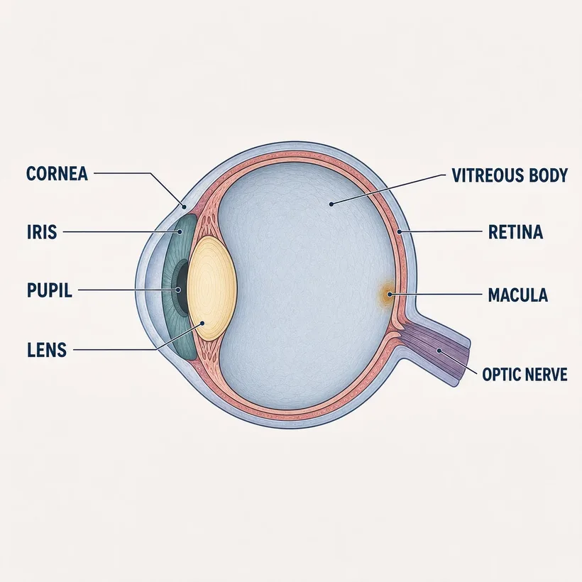
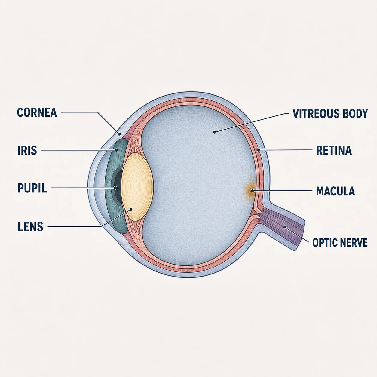
Light passes cornea → pupil → lens → vitreous → retina; the optic nerve carries the picture.
The structures
- Cornea — Clear front window — most of the eye's focusing power
- Iris & pupil — Aperture — CN III controls constriction
- Lens — Fine focus; clouds into cataract
- Vitreous body — Gel filling the globe
- Retina — Light → nerve signal; detaches painlessly
- Macula & fovea — Central, sharp vision
- Optic nerve (CN II) — Signal to the brain; swells with high ICP (papilledema)
Why it matters on shift
- A blown (fixed, dilated) pupil on one side = CN III compression = neuro emergency
- Sudden painless vision loss or 'curtain coming down' = retinal detachment or artery occlusion — minutes matter
- Painful red eye + halos + nausea = acute angle-closure glaucoma, not conjunctivitis
Source: Anatomical structures per OpenStax Anatomy & Physiology, 2nd ed. (openstax.org, CC BY 4.0). Educational reference — verify clinical application against your facility's policy.Reference only — verify against your facility's policy.